
Types of amyloidosis
AL amyloidosis
AL amyloidosis is one of the main forms of systemic amyloidosis. It is an acquired disease caused by abnormal plasma cells in the bone marrow that produce misfolded immunoglobulin light chains. These light chains can form amyloid deposits in different organs. Because organ damage may progress rapidly, early diagnosis and specialist care are essential.

01
What is AL amyloidosis?
Key idea
AL amyloidosis is not passed down through families, and it is not a disease of the organs it damages. It starts with a small group of cells in the bone marrow.
An acquired disease
The name says what it is: Amyloid made of immunoglobulin Light chains. Abnormal monoclonal antibody fragments build up in the body as insoluble fibrils, and as they accumulate they progressively stop organs from working normally.
Unlike hereditary forms of amyloidosis, AL is not inherited and cannot be passed on to your children. These deposits can affect every organ in the body except the brain.
Systemic or localised
In the systemic form — by far the most common and the subject of this page — light chains circulate in the blood and deposit across several organs at once.
In localised forms, the cells producing the light chains sit in one place and the deposits stay confined to a single site: the conjunctiva, larynx, airways, bladder or digestive tract. The outlook is generally much better.
02
How AL amyloidosis develops
From a normal cell in the bone marrow to deposits in the organs — the four steps that turn a routine biological process into a disease.
Step 01
Plasma cells
Plasma cells and B lymphocytes in the bone marrow produce antibodies. Each antibody is built from two heavy chains and two light chains.
Step 02
A clone appears
Occasionally a single cell becomes, in effect, immortal. It multiplies into a clone that produces one identical monoclonal antibody, over and over.
Step 03
Light chains misfold
In roughly 1% of cases these free light chains are unstable. They stack together into amyloid fibrils that the body cannot break down.
Step 04
Organ deposits
The fibrils settle in the tissues, stiffening them and disrupting their function. Which organs are hit — and how hard — shapes the whole clinical picture.
Estimated incidence: approximately 8–12 new cases per million people per year. Reliable worldwide figures are limited, and underdiagnosis is likely.
Most often diagnosed in older adults, commonly between 60 and 70 years of age.
~1%
of monoclonal light chains form amyloid fibrils
Related conditions you may have heard of
MGUS — monoclonal gammopathy of undetermined significance
A monoclonal immunoglobulin is present, but it involves fewer than 10% of the cells in the bone marrow. MGUS becomes more common with age: it is found in about 5% of people at 60, and in up to 10% of people over 80. Most people with MGUS never develop amyloidosis.
Myeloma
Myeloma involves more than 10% of bone marrow cells. When myeloma is found alongside AL amyloidosis, it is typically indolent — slow-moving. The two conditions come from the same family of cells, which is why AL is treated with regimens originally developed for myeloma.
03
Symptoms
Symptoms depend entirely on which organs the fibrils have reached. Because each of them is common on its own, AL amyloidosis is easily mistaken for something more ordinary — which is why diagnosis is so often delayed.
> 2 in 3
Kidneys
The most frequently affected
Amyloid damages the glomerular filter, letting albumin escape into the urine. This causes swelling of the legs or more widespread oedema, and a decline in kidney function that can eventually require dialysis.
~60%
Heart
The most serious
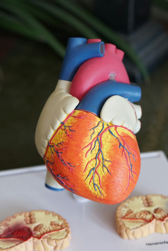
The heart muscle thickens and loses its ability to relax and fill — a restrictive cardiomyopathy. It brings fatigue and breathlessness, at first on exertion and later at rest. Abnormal rhythms may require a pacemaker.
~20%
Nerves
Peripheral and autonomic
Abnormal sensations begin in the feet and move up to the legs, thighs and hands. When the autonomic nerves are involved, they cause digestive trouble and a drop in blood pressure on standing, with fainting and falls.
Digestive tract
Diarrhoea and constipation, often alternating, and sometimes bleeding. Deposits in the tongue make it enlarge and can alter the sense of taste.
Skin
Amyloid weakens the small blood vessels of the skin, so it bleeds and bruises easily. Bruising around the eyes is a particularly telling sign.
When to seek advice
No single sign below points to AL amyloidosis on its own. Several of them together, especially if they are new and unexplained, are worth raising with a doctor.
- Swelling of the ankles and legs
- Fatigue that rest does not relieve
- Shortness of breath on light exertion
- Numbness, tingling or pain in the hands and feet
- Diarrhoea alternating with constipation
- Unexplained weight loss
- An enlarged tongue
- Bruising, especially around the eyes
- An irregular heartbeat
AL amyloidosis can become life-threatening quickly. Catching it early is crucial.
04
How AL amyloidosis is diagnosed
Reaching a diagnosis means answering three separate questions: is there amyloid, what kind of amyloid is it, and how much damage has it already done? Some of these tests may sound daunting. Most are quick, and your team will talk you through each one.
01
Confirm the amyloid
Diagnosis always requires a tissue biopsy. Stained with Congo red, amyloid deposits reveal themselves under polarised light.
02
Type the amyloid
The deposits are then typed to separate AL from the other forms, in particular the hereditary ones. This step is critical: the treatments have nothing in common.
03
Characterise the blood disorder
A bone marrow sample identifies the cells producing the abnormal antibody, while blood and urine tests measure the free light chains themselves.
04
Assess organ involvement
Heart: echocardiography, cardiac MRI where indicated, biomarkers (BNP, NT-proBNP, troponin), ECG and 24-hour Holter monitoring.
Kidneys and liver: creatinine, albumin, proteinuria and liver function tests.
Imaging: abdominal ultrasound or CT.

05
Treatment
Treatment does not remove the deposits. It shuts off the supply — destroying the cells that produce the toxic light chains, so the body can begin to clear what has built up.

Treatment targets the abnormal plasma-cell clone in the bone marrow to rapidly reduce the production of amyloid-forming light chains. The regimen and its intensity are tailored to the person’s organ involvement, general health and treatment tolerance. Response is monitored with blood tests, while recovery or progression of organ involvement is assessed separately over time.
First line: Dara-VCd
The most frequently used combination today: a monoclonal antibody added to a proteasome inhibitor, an alkylating agent and a corticosteroid.
Proteasome inhibitors
These agents push the abnormal plasma cells into programmed cell death. In use for well over a decade, they substantially reduce their number.
Immunomodulators
These agents slow the multiplication of the abnormal cells rather than killing them outright.
Monoclonal antibodies
They recognise the plasma cells producing the harmful light chains and mark them for destruction.
Corticosteroids
Steroids are not given alone. They are always paired with one of the agents above, whose effect they amplify.
Emerging therapies
Targeted agents and bispecific antibodies — a newer class — are being used alone or in combination. Trials continue; ask your team what is open to you.
Autologous stem cell transplant
In selected patients, high-dose chemotherapy followed by autologous stem cell transplantation may be considered in an experienced specialist centre. Eligibility depends on organ involvement, overall fitness and treatment risk. This approach is not suitable for most patients.
Supportive care runs alongside
Supportive care is provided alongside treatment of the plasma-cell disorder. It aims to manage symptoms and complications affecting the heart, kidneys, nerves and other organs. The measures used depend on the organs involved, the severity of disease and the resources available locally.
06
Follow-up and outlook
Monitoring
During and after treatment, blood tests are used to monitor the abnormal light chains, treatment safety and organ function. The frequency of follow-up and the decision to restart treatment depend on the individual situation and should be determined by the specialist team.
Outlook
The outlook varies widely and is strongly influenced by the extent of heart and other organ involvement at diagnosis, the depth and speed of the haematological response, and the person’s overall health. Outcomes have improved substantially with earlier diagnosis, more effective plasma-cell directed treatment and specialist supportive care. However, advanced cardiac involvement remains a major risk factor.
Of everything that shapes the outcome, the delay before diagnosis is the one we can still change.
FAQ
Common questions
Is AL amyloidosis inherited?
No. Unlike several other forms of amyloidosis, AL is acquired during life, not carried in your genes. It cannot be passed on to your children, and your relatives are not at increased risk because of your diagnosis.
Can AL amyloidosis affect the brain?
No. Amyloid light-chain deposits can reach every organ in the body except the brain. Nerves outside the brain and spinal cord, however, are affected in around one patient in five.
Is AL amyloidosis a cancer?
It begins with a clonal disorder of the plasma cells in the bone marrow — the same family of cells involved in myeloma — and it is treated by haematologists using myeloma protocols. But it behaves differently, and where myeloma accompanies amyloidosis it is usually indolent. Your haematologist is best placed to explain what this means in your own case.
What are free light chains?
Antibodies are built from two heavy chains and two light chains. When light chains are produced in excess by a clone of plasma cells, they circulate in the blood on their own — free. In AL amyloidosis these free light chains are the raw material of the deposits, which is why measuring them tells your team whether treatment is working.
What is the difference between AL and ATTR amyloidosis?
They differ in the protein that misfolds. In AL it is an immunoglobulin light chain made in the bone marrow, and the disease is acquired. In ATTR it is transthyretin, a protein made by the liver; that form may be inherited (hATTR) or come with age (wtATTR). Because the source proteins differ, so do the treatments — which is why typing the amyloid is such an important step.
This page is intended as general information for patients, families and healthcare professionals. It does not replace medical advice. Always discuss your situation with your own specialist team.