
Other types
Rare hereditary amyloidosis
Beyond transthyretin, a handful of other inherited proteins can misfold and form amyloid. These forms are extremely rare, they share no clinical signature of their own, and the kidneys bear the brunt. Naming the exact protein is not an academic detail: it is what decides the treatment.
01
What are they?
Key idea
These forms cannot be recognised at the bedside. They have no clinical signature of their own. Only naming the protein tells the doctor what to do.
Beyond transthyretin
Alongside the amyloidoses of transthyretin, several other hereditary forms arise from mutations in different proteins altogether. Each mutation alters the genetic code so that a protein folds wrongly, and the misfolded protein deposits as amyloid.
Transthyretin remains by far the most frequent of the hereditary forms. The others described on this page are extremely rare.
No signature of their own
They present with no distinctive clinical features. Nothing in the symptoms points to one protein rather than another — which is why they are so easily missed, and why the laboratory has the last word.
The kidneys are the principal target. Which other organs are involved depends entirely on which protein has mutated.
02
The proteins involved
All are transmitted in an autosomal dominant way. Each has its own tropism — and, crucially, its own treatment.
AFib
Fibrinogen A-alpha chain mutation. An exclusively renal disease, and one of the more common hereditary forms in Western Europe. The protein is made by the liver — which opens a therapeutic door the other forms do not have.
AApoAI
Apolipoprotein A-I mutation. Affects the kidneys, the liver and the peripheral nerves. Several members of the apolipoprotein family can be implicated — A-I, A-II, A-IV, C-II and C-III.
ALys
Lysozyme mutation. Renal involvement in the first place, with deposits in the liver as well. The protein is produced by several tissues, not by the liver alone — a fact that changes everything about its treatment.
AGel
Gelsolin mutation. Principally causes cranial neuropathy and corneal lattice dystrophy. Most prevalent in Finland.
03
Symptoms
For a long stretch there is nothing at all to feel, while the deposits quietly accumulate. The symptoms, when they come, are those of a kidney that has been failing for years.
The kidneys
The primary target
The early phase is silent: amyloid settles in the kidney without announcing itself. Later come swelling of the ankles, high blood pressure, fatigue, pallor and breathlessness — the general symptoms of advanced kidney failure.
Other organs
It depends on the protein
Beyond the kidneys, the pattern shifts from one mutation to the next: the liver, the peripheral nerves, the cornea. Two people with the same symptoms may have two different diseases.
What the tests show
Before the symptoms
Protein spilling into the urine — proteinuria — is often the first abnormal finding. A rising creatinine in the blood signals that the kidney is no longer filtering as it should.

When to seek advice
None of these signs is specific to amyloidosis. What should raise the question is their combination with a family history of the disease.
- Swelling of the ankles
- High blood pressure
- Fatigue and pallor
- Shortness of breath
- Protein found in the urine
- A rising creatinine on blood tests
- Amyloidosis in a parent, a sibling or a child
For a long time there is nothing to feel. The deposits accumulate in the kidney in silence, and the first sign is often a laboratory result.
04
How they are diagnosed
Since nothing in the clinical picture distinguishes one of these forms from another, the whole diagnosis rests on the laboratory — and, at the end, on a gene.
01
A family history
Amyloidosis among parents, siblings or children. Several affected members point towards a genetic form — though a single, isolated case remains entirely possible.
02
Find the amyloid
A biopsy from an accessible site — the salivary glands, the digestive tract. More often than not it is a kidney biopsy, taken without amyloidosis being suspected at all, that uncovers it. Congo red staining confirms the deposits.
03
Type it
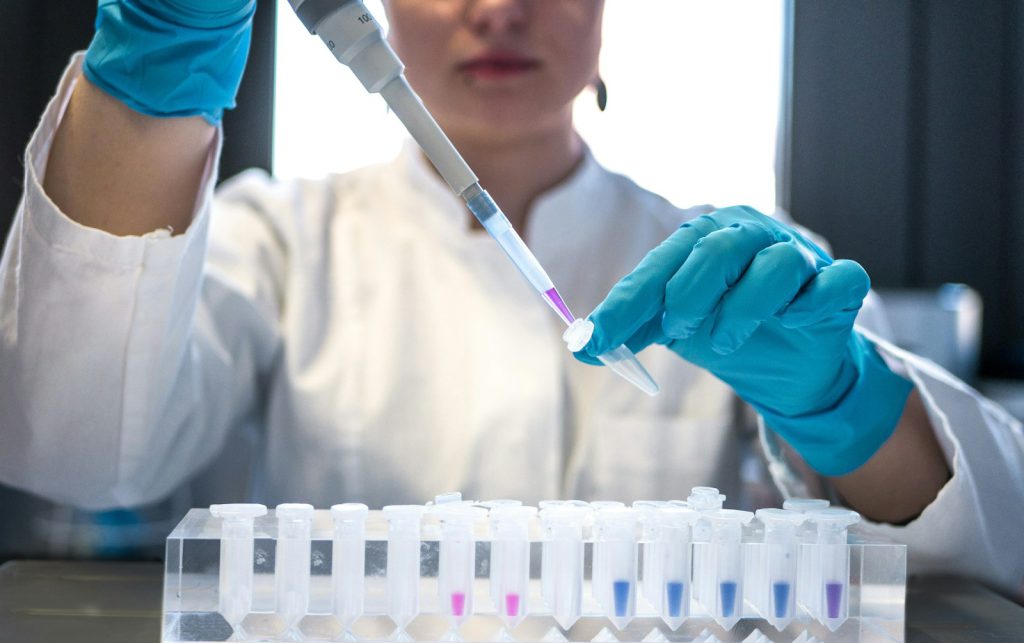
Immunohistochemistry, using antibodies specific to each protein, narrows down which one has been deposited.
04
The genetic test
This is the definitive method. It names the protein, and in naming it, it names the treatment.
05
Treatment
Here is why the precise diagnosis matters so much.
The symptoms may be identical from one rare form to another, while the treatments are entirely different. Establishing exactly which one it is changes what can be done.
Stop the production at its source
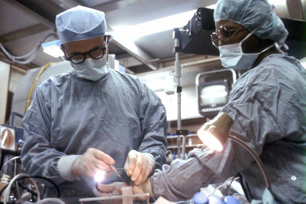
The definitive treatment consists of eliminating the production of the mutated protein. That is possible only when the protein is made by the liver — because a liver can be replaced.
The fibrinogen alpha chain is made by the liver, so a liver transplant addresses the cause. Lysozyme is produced by several tissues at once, and no transplant can remove them all. Same kidney failure, same appearance under the microscope, opposite conclusions.
Liver transplant
Possible where the mutated protein is made by the liver, as it is for the fibrinogen alpha chain. Not an option for lysozyme, which several tissues produce.
Dialysis
When the kidneys can no longer do their work, dialysis takes over. It sustains, but it does not touch the cause.
Kidney transplant
Often possible, and often transformative. But it replaces the organ, not the source: the disease can return in the transplanted kidney.

06
Follow-up
Watching the kidneys
Follow-up is built around kidney function. It is measured, and measured again, because it is the thing that decides how the disease will unfold and when a transplant becomes necessary.
An unpredictable course
The way these diseases progress cannot be foreseen. Kidney function may decline slowly over many years, or rapidly. That unpredictability is precisely why surveillance is continuous rather than occasional.
Their rarity is the difficulty. Naming the protein is what turns a disease nobody can treat into one that can be.
FAQ
Common questions
Are these forms inherited?
Yes. They are transmitted in an autosomal dominant way, like hereditary ATTR. A mutation in one parent is enough. That said, an isolated case in a family with no known history is entirely possible.
Why does the exact protein matter so much?
Because the treatments have almost nothing in common, while the symptoms may be indistinguishable. A liver transplant addresses the cause of the fibrinogen form and can do nothing for the lysozyme form. Two patients with the same failing kidneys need two different answers, and only the laboratory can say which.
Can I be tested?
A genetic test exists and is the definitive method. In France it is regulated: adults only, preceded by interviews with a psychologist, with results returned after several weeks. Screening usually starts with siblings and adult children. The rules differ between countries — your centre will tell you what applies where you live.
Which organ is affected?
The kidney, principally, in all of these forms. Beyond that it depends on the protein: the liver, the peripheral nerves, or the cornea may also be involved. There is no combination of symptoms that identifies one form rather than another.
Does a kidney transplant cure the disease?
No. It replaces the organ that has failed, not the protein that destroyed it, and the disease can recur in the transplanted kidney. Where the mutated protein is made by the liver, a liver transplant does address the cause — which is another reason the exact diagnosis is worth the wait.
This page is intended as general information for patients, families and healthcare professionals. It does not replace medical advice. Genetic testing pathways, transplant eligibility and access to specialist treatments vary between countries. Always discuss your situation with your own specialist team.