
Other types
Localised amyloidosis
Localised amyloidosis affects one organ, and only one. The plasma cells that make the abnormal light chains sit inside that organ; nothing travels through the blood, so nothing is deposited anywhere else. Among the amyloidoses, which are almost always multisystemic, it is the exception.
01
What is it?
Key idea
In localised amyloidosis, amyloid deposits form and remain within a single organ or tissue. The precursor protein and the cells producing it vary according to the affected site and subtype.
One organ, and only one
Localised amyloidoses affect a single organ or tissue. The great majority of them are AL amyloidoses — deposits made of immunoglobulin light chains, exactly as in the systemic form.
What sets them apart is not the protein. It is where the light chains come from, and where they go.
A clone that stays put
The plasma cells producing them proliferate inside the organ itself. The light chains never pass into the bloodstream, and so they are never carried anywhere else. The deposits form where they are made, and nowhere beyond.
Most amyloidoses are multisystemic. Localised forms are the exception, and this is the mechanism that makes them so.
02
Localised or systemic?
Two diseases built from the same protein. Everything that matters — the organs at risk, the treatment, the outlook — turns on which of the two it is.
LOCALISED
Confined to one organ
The plasma cells proliferate on the spot, and the light chains they make stay where they are made. Nothing enters the circulation, so no distant organ is ever reached.
The treatment is local: surgery, radiotherapy, or a treatment applied directly to the organ.
SYSTEMIC (AL)
Carried through the blood
A clone of plasma cells in the bone marrow releases light chains into the bloodstream. They deposit in the heart, the kidneys, the nerves — wherever the blood takes them.
The treatment is chemotherapy, aimed at the marrow. Read about AL amyloidosis →
The whole purpose of the diagnostic work-up is to establish which of the two you have. Under the microscope the deposits look alike. The diseases do not.
03
Where it occurs
Since only one organ is involved, the symptoms are entirely those of that organ. They say nothing about amyloidosis, and everything about where the deposits sit.
The airways
Larynx, trachea, bronchi
The voice changes, little by little, over months — a dysphonia that is often the first thing anyone notices. A cough settles in, and breathing becomes harder.
The lower urinary tract
Bladder, ureter
Blood appears in the urine. Passing water becomes difficult, and pain settles low in the abdomen. These are the signs that usually bring the patient to a urologist.
Other sites
Less commonly
Deposits may also stay confined to the conjunctiva of the eye, to the digestive tract, or to the skin. Wherever a small group of plasma cells has settled, amyloid can accumulate.

When to seek advice
Each of these has a great many ordinary explanations, and amyloidosis is almost never the first one considered. What matters is that a symptom which persists, and which nothing accounts for, deserves to be looked at properly.
- A voice that changes over months
- A cough that does not settle
- Breathlessness
- Blood in the urine
- Difficulty passing urine
- Pain low in the abdomen
These are exceptional diseases, and they are rarely suspected at the outset. More often than not, the biopsy is taken to rule out a tumour.
04
How it is diagnosed
The diagnosis is usually made by accident: a biopsy taken because a tumour was suspected comes back showing amyloid instead. What follows is harder, and more important — proving that the disease stops there.
01
A biopsy of the organ
A sample is taken from the affected site, and it reveals amyloid deposits where a malignancy had been suspected. This is very often how these diseases come to light.
02
Type the deposits
Immunohistochemistry can establish which type of amyloid is involved, although in practice precise typing is carried out infrequently.
03
Prove that it is localised
This is the critical objective: to affirm the localised character of the lesions, and to make sure there is no amyloid in any other organ. It rests on clinical examination together with further investigations — and it is what separates this disease from systemic AL amyloidosis.
05
Treatment
Treat the organ, not the marrow. There is no single protocol here — the approach is decided case by case.
Guided by what the organ can no longer do
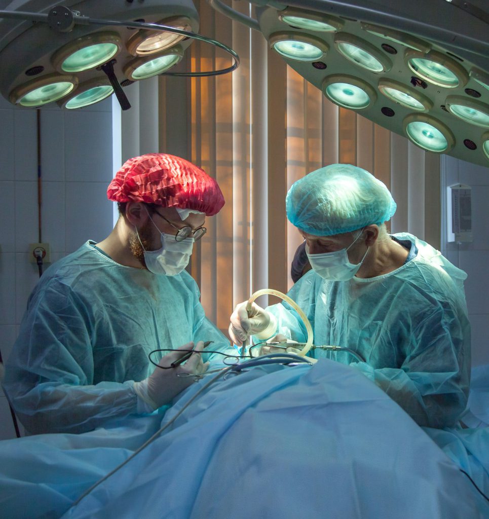
Management varies from one patient to the next, and what guides it is how far the deposits have compromised the organ’s function. A voice that has become hard to use, a bladder that no longer empties — these, rather than the size of the deposit, decide what is done.
Because the light chains never leave the organ, there is nothing in the bloodstream for a systemic drug to act upon. That single fact is why the treatment stays local.
Surgical removal
The deposits can be taken out surgically where they interfere with the way the organ works. It is the most direct answer to a problem that sits in one place.
Radiotherapy
For selected lesions, radiotherapy is used instead — where surgery would cost more than it gives back.
Local treatment
For amyloidosis of the bladder, a medical treatment applied locally is possible — delivered to the organ rather than to the whole body.
When is systemic treatment used?
Systemic treatment is usually not required once the amyloid type has been confirmed and systemic disease has been excluded. Treatment should be tailored to the affected organ and the individual patient.
Where it is considered, the decision belongs to a specialist team that has first established, beyond doubt, that the disease is truly confined to one organ.

06
Follow-up
Two kinds of watching
The first is local, and belongs to the specialist of the organ concerned — an ENT surgeon where the larynx is involved, a urologist where the bladder is. They watch the deposit itself, and what it does to the organ.
Why the second matters
The second is general, and its purpose is to verify that the disease has remained localised. The localised character of an amyloidosis is not something established once and then filed away. It is something checked, and checked again.
It is the same protein as in AL amyloidosis. It is not the same disease — and the whole difference is measured in where the light chains go.
FAQ
Common questions
Is localised amyloidosis the same as AL amyloidosis?
The deposits are made of the same protein — immunoglobulin light chains — and they look alike under the microscope. But in the localised form the plasma cells producing them sit inside the affected organ, and the light chains never enter the bloodstream. No other organ is reached. The mechanism differs, the treatment differs, and so does what the diagnosis means for you.
Can it become systemic later on?
The general follow-up exists precisely to answer that question, over time. Alongside the local monitoring of the organ, a broader surveillance verifies that the disease has stayed where it was found. It is not a formality: the localised character is confirmed by checking, not assumed.
Will I need chemotherapy?
Rarely. Systemic medical treatment is seldom indicated here, because there is no marrow clone to suppress and no circulating light chain to bring down. Treatment is directed at the organ — surgery, radiotherapy, or a locally applied medical treatment in the case of the bladder.
Which organs can be affected?
Most often the airways — larynx, trachea, bronchi — or the lower urinary tract, meaning the bladder and the ureter. Deposits may also remain confined to the conjunctiva of the eye, to the digestive tract, or to the skin.
Is the outlook better than in systemic amyloidosis?
Generally, yes — considerably so. Because the deposits stay in one place, the vital organs that systemic amyloidosis threatens are not involved. That said, these are exceptional diseases and each situation is its own; what the deposits do to the organ they occupy is what your specialist team will discuss with you.
This page is intended as general information for patients, families and healthcare professionals. It does not replace medical advice. Always discuss your situation with your own specialist team.